
PET CT Scan in Israel
 A PET/CT scan is one of the most current diagnostic tools available at present to physicians. Combining positron-emission and computed tomography capabilities it can map out changes in tissue structure. A built-in computer measures the intensity of contrast uptake. These factors help specialists evaluate the structure and condition of a patient’s internal organs.
Please note: If you have PET/CT results you can request a second opinion from an Israeli radiologist.
How Does a PET/CT Scan Work?
PET (positron-emission tomography) is a non-invasive nuclear medicine technique, where images are obtained using specialized radioactive tracers. It is used to evaluate physiolological and biochemical processes within the body. CT (and MRI) scans, on the other hand, assess structural changes.
Biochemical process disruptions, which often precede anatomic damage, are seen with any serious condition. A PET scan helps enhance the diagnostic process with data about the lesions/foci, increasing diagnostic accuracy.
18F-FDG (Fludeoxyglucose) is the most common radio-tracer employed. The agent’s properties lead to its abnormally high uptake in pathologic foci (primarily in malignant foci / mets), making it useful in assessing energy metabolism and an invaluable tool in diagnosing oncological diseases.
Still, a PET scan has a significant drawback: It does not provide any data about an organ’s structure, which makes determining the exact location of the pathological foci problematic. To overcome this device an innovative PET/CT scanner was developed. It allows two investigations to be performed at once. The resulting combined image shows a detailed anatomical map with metabolic processes superimposed.
The scans are predominantly used to diagnose and monitor patients with cancer and heart conditions.
A PET/CT scan may be indicated in the following cases:
- Evaluating metastases
- Ruling out cancer recurrence
- Determining where a tumour is malignant or benign
- Diagnostics and evaluation of coronary artery disease / risk of CAD
- Pre-operative evaluation (heart surgeries)
- Evaluation post heart surgery
- Monitoring response to treatment
- In neurology PET scans can be used to diagnose dementia, Alzheimer’s, Parkinson’s and Huntington’s diseases
PE/CT Scan: Stages
The radiotracer will be administered approximately 2 hours prior to the scan (the waiting period is needed for it to spread all over your body). Before the scan you will be asked to lie on a specialized bed, which will be moving through the scanner during the investigation. The procedure will take approximately 45 minutes.
The radiotracer can either be given intravenously (chills are common side effect), or perorally (it has no taste or smell). It is evacuated from the body naturally within several days. As the dosage is very small, you have nothing to worry about.
Please notify your treating physician in the following cases:
- You are or may be pregnant or breastfeeding
- If you take any medications on a regular basis (including vitamins)
- If you have any allergies
- If you have been diagnosed with diabetes (special preparation will be needed in such event)
You will be asked to fast for at least 6 hours prior to the test as the presence of food in your organism may lead to incorrect interpretation of the results. You are allowed to keep drinking water, but please refrain from high-calorie beverages / soft drinks.
A PET/CT scan is usually painless and complications-free. If a scan of the pelvic region is required a catheter may need to be inserted into your bladder. This is known to cause some discomfort. You will be asked to refrain from moving during the scan. Once the investigation is complete you can resume your normal activities (unless specified otherwise). In order to expedite evacuation of the radiotracer you’re advised to drink more water than you usually do.
PET/CT Diagnostic at Assuta Medical Center
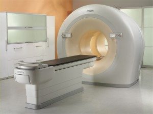
 Israel’s Assuta Clinic utilizes the most advanced PET/CT scanners built by Phillips. These are the first systems available that can combine cutting-edge radiologic oncology techniques, increase diagnostics accuracy, provide maximum comfort to patients and obtain images of the highest quality. Phillips utilized the ground-breaking Time-of-Flight technology to improve scan quality. Specialized software helped shorten the time of the scan. The scanner’s increased sensitivity allows reduced radiotracer dosages.
Please not that any PET/CT scan performed at Assuta utilizing the Time-of-Flight technology is interpreted twice: by a radiologist and a specialist in nuclear medicine.

